DEEP VENOUS THROMBOSIS (DVT)
vascular.co.nz>deep venous thrombosis
What is a
Deep Vein or Venous Thrombosis (DVT)?
What are the symptoms of a Deep Venous Thrombosis?
What is the economy class syndrome?
Does Deep Venous
Thrombosis occur at other times?
Risk Factors for Deep Venous Thrombosis
Why is deep Venous
Thrombosis important?
Can the economy class syndrome
(DVT) be prevented?
How is Deep Venous Thrombosis diagnosed?
Can Deep Venous Thrombosis be treated?
Other treatments for DVT
Are there long term complications
of Deep Venous Thrombosis?
Latest news
References
Deep Venous Thrombosis links
What is a deep vein or venous thrombosis (DVT)?
There are two main groups of veins in the legs: the superficial veins and the deep veins. When blood clots form in the deep veins, this is known as a Deep Vein Thrombosis or Deep Venous Thrombosis (DVT). These terms mean the same. The deep veins cannot be seen externally. The superficial veins can be seen through the skin and sometimes form varicose veins when they become enlarged. Superficial veins can sometimes develop blood clots if phlebitis develops (superficial thrombophlebitis). Thrombosis can actually take place in any of the veins throughout the body, but is most frequent in the legs.
DVT in the legs can be divided into 3 main types classified by their location. Firstly ilio-femoral DVT affects the major vein in the pelvis (iliac vein) draining the leg and may also involve the femoral vein in the thigh. Femoro-popliteal vein DVT predominantly affects the femoral vein in the thigh and the popliteal vein behind the knee. Calf vein (tibial vein) DVT predominantly affects the smaller deep veins in the lower leg. These divisions are largely arbitrary and DVT may affect all of these veins or segments of a vein that don't readily fit into this classification. The divisions are useful in terms of assessing severity and risk of complications with ilio-femoral DVTs likely to present with the most severe symptoms and highest risk of subsequent complications and calf vein DVT with the lowest risk..
What are the symptoms of a deep venous thrombosis?
Many patients may have minimal or no symptoms and this is known as a silent DVT. These patients may never know they have had a DVT.
The main symptoms of a DVT are pain and swelling in the affected leg, particularly in the calf. The calf may be slightly red and tender. These symptoms can vary widely in their severity and are also not very specific. It can be difficult to make the diagnosis of DVT on the symptoms alone, because many other unrelated disorders can also cause similar symptoms.
In mobile patients who present with symptoms suspicious of a DVT, 75% (3 of every 4 patients) will not have a DVT (Fancher TL et al, 2004).
| High risk groups for DVT |
|---|
| Previous DVT |
| Previous pulmonary embolus |
| The elderly |
| Malignant disease |
| Extensive trauma/extensive surgery |
| Obesity |
| Abdominal or pelvic surgery rather than upper limb or head and neck surgery |
| Contraceptive pill |
| Congestive heart failure. Myocardial infarction |
| Underlying thrombophilia |
Occasionally DVT can be very severe and affect the deep and superficial veins. In these circumstances the venous drainage from the limb may be so impaired that venous gangrene can develop. There is often an underlying cause for this event, such as an underlying cancer. It can be a very difficult situation to treat.
What is the economy class syndrome?
The development of a DVT following a long flight has been called the economy class syndrome (Cruikshank JM et al, 1988). The first such recorded event was in 1946 on a non-stop flight lasting 14 hours (Homans J, 1954). The economy class syndrome has now expanded to refer to any flight related DVT especially when travelling economy class because of the more limited leg room.
Does deep venous thrombosis occur at other times?
DVT is not only associated with air travel but has been recorded after any long period of immobility, such as a long car or rail journey or more commonly during a period in hospital, particularly in severely ill patients during prolonged periods of immobility. It is rare following air travel and much more commonly occurs in patients who have not been on a recent flight.
Deep venous thrombosis can occur in 20% of patients undergoing major surgery and over 40% of patients undergoing major orthopaedic (bone) surgery.
Risk factors for deep venous thrombosis
It is thought that a combination of factors lead to the development of a DVT. Some of these factors can be described as external factors. For instance a combination of immobility and cramped seats, with the front of the seat pressing on the calf, may be important in the economy class syndrome. Aircraft cabin pressure may also play a role. There do seem to be factors related partcularly to air travel which cause activation of the coagulation system (Schreijer AJM et al, 2006). A UK government report (Sunday Times, September 2nd, 2001) has investigated factors which could influence the development of DVT during air travel. It has indicated that decreased cabin pressure and altered sleep patterns because of jet lag may be important in the development of DVT. They have suggested that more studies to investigate the links between air travel and DVT should be funded.
Immobility is also important in patients undergoing surgery. The combination of a long surgical operation and a prolonged period of bedrest will increase the risk of DVT.
Apart from these external factors there may also be internal factors which increase the likelihood of an individual person developing a DVT. Important risk factors include age over 40 years, pregnancy, presence of cancer, hormone therapy (hormone replacement therapy, the oral contraceptive pill) and dehydration.
If you have had a previous DVT then you are always at a slightly increased risk of a further DVT, particularly soon after your first DVT. This risk does decrease with time, but can be as high as 25% at 4 years. The risk of recurrence appears to be less in patients who develop a post-operative DVT.
If you have had a previous DVT or have blood disorders that make clotting more likely (thrombophilias) then they also put you at increased risk. At least 30% of patients with DVT and no obvious cause will have thrombophilia on testing. Factor V Leiden is the commonest abnormality and found in 12-30% of patients who have a DVT.
May-Thurner syndrome is a poorly recognised cause of left sided ilio-femoral DVT. It is a condition in which the left common iliac vein in the pelvis is compressed by the right common iliac artery which passes in front. Elegant studies using magnetic resonance venography have confirmed the association with ilio-femoral DVT (Fraser DGW et al, 2004). They have also shown that thrombus (clot) is less likely to resolve and the vein to re-open in the presence of this syndrome (Fraser DGW et al, 2004).
Thrombosis is important to recognise because it may be dangerous in two ways.
Firstly, if a piece of blood clot breaks off from the veins in the legs and travels along the veins to the lungs (pulmonary embolism, PE) it can be fatal in a small number of people. Even if it is not fatal such a blood clot can make you very seriously ill. Pulmonary embolism causes symptoms such as shortness of breath, chest pain, cough, coughing up blood (haemoptysis), and collapse. Most patients with PE have no leg symptoms at diagnosis (Blann AD, Lip GYH, 2006).
Secondly, after a clot has formed in the legs it can damage the valves in the veins or block the veins. In either case this may cause problems in the legs in the future (see chronic venous insufficiency). It has been estimated that up to 90% of patients with ilio-femoral DVT will subsequently develop signs and symptoms of chronic venous insufficiency with up to 15% developing venous ulcers. As the blood clot clears from the leg and venous blood flow resumes this is abnormal with reflux or reverse flow developing and increasing with time.
Can the economy class syndrome (DVT) be prevented?
For the majority of people going about their normal business there is no need to take any special precautions to reduce the risk of DVT. Prevention is important if you are known to be at high risk of DVT or are to undertake an activity which could put you at increased risk.
DVT can be prevented by taking sensible precautions. The most important preventive measure is activity. Standing up, stretching and taking a brief walk every hour during air travel will help to reduce your risk. It is important not to become dehydrated - so drink water or soft drinks regularly. This will also aid mobility!
It is important to exercise the calves of the legs when sitting. The calves can be gently exercised when sitting by pressing the front of the feet onto the floor and moving the heels up and down off the floor.
Graduated compression stockings are particular types of stocking that provide maximum compression around the ankle area. The compressive effect then becomes less further up the leg. This improves venous flow in the deep veins. Compression stockings are proven in hospital studies to reduce the risk of DVT. It is important that the stockings are fitted correctly.
If you are at particular risk of DVT, then it may be sensible to take 150mgs of aspirin before your flight. Aspirin can cause irritation of the stomach and can rarely lead to bleeding from the stomach. It is important not to take aspirin, unless you know you are safe to do so and have had no previous problems with vomiting blood or peptic ulcers.
Many patients undergoing surgery have injections of low dose (prophylactic) heparin under the skin once or twice per day to reduce the risk of blood clots forming. These patients can also have pneumatic cuffs fitted to the calfs or feet which intermittently inflate and deflate throughout surgery. This encourages the flow of blood in the veins of the legs, and helps to prevent long periods where the blood in the veins is slow moving and may lead to thrombosis. There are detailed guidelines available in Australia and New Zealand to assist in using methods to prevent post-operative thrombosis.
A recent randomised trial (Scurr et al 2001) has produced some unexpected results. Volunteers were allocated to wearing or not wearing below knee stockings during long flights, returning within 6 weeks. Scanning of the veins and blood tests were performed within 48 hours of return to detect DVT. In the group not wearing stockings a high incidence of silent DVT was detected (1 in 10). Volunteers wearing stockings did not suffer any DVTs. This study has been criticised on various grounds, especially the fact that DVT was so common in the group not wearing stockings. All the DVTs detected were very small and present in the calf veins which is an area where the ultrasound scan can have difficulty being accurate. Despite this it is clear that this finding requires further detailed studies to either confirm or refute the findings.
How is Deep Venous Thrombosis diagnosed?
The diagnosis of DVT may be suspected by the symptoms and signs in an individual patient and the circumstances of the patient. For instance, pain and swelling in one calf, in a patient after major surgery or a long flight, will raise a suspicion of DVT, which will require exclusion with further tests. The same symptoms that develop following a game of squash are more likely to be due to a muscle injury. Clinical diagnosis alone is unreliable and inaccurate and further tests are required.
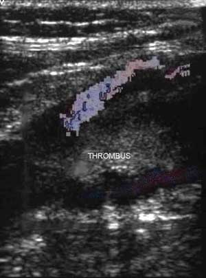 The main test used to exclude or
diagnose DVT is
Duplex ultrasound scanning. This is a simple, painless test with a
high degree of accuracy. Ultrasound can demonstrate clot within the deep veins.
It is particularly accurate in the larger veins of the leg. The image on
the left is an ultrasound scan showing thrombus (the blood clot) with some blood
flowing around the clot.
The main test used to exclude or
diagnose DVT is
Duplex ultrasound scanning. This is a simple, painless test with a
high degree of accuracy. Ultrasound can demonstrate clot within the deep veins.
It is particularly accurate in the larger veins of the leg. The image on
the left is an ultrasound scan showing thrombus (the blood clot) with some blood
flowing around the clot.
In the calf veins ultrasound is more difficult, but can be accurate in experienced hands. Unfortunately ultrasound can be time consuming and costly. As the majority of tests will be normal, clinicians are trying to find ways to reduce the number of normal scans performed. One way to do this is to measure D-dimer levels.
Measurement of D-dimer (a marker of coagulation or blood clotting) in blood is gaining increasing popularity as a rapid and inexpensive screening test. This test is especially useful for excluding DVT if the results are normal. In a patient at low or moderate risk, a normal D-dimer test can safely rule out DVT (Fancher TL, 2004). If the D-dimer is abnormal, then confirmation of DVT using Duplex scanning is important. This is because other situations where blood clotting occurs can lead to an increase in D-dimer levels. No test is foolproof, and in a patient with a high risk of DVT and a normal D-dimer Duplex scanning should be performed.
Other tests such as venography and plethysmography are much less commonly used today, but venography is the most accurate test in the diagnosis of DVT.
Can deep venous thrombosis be treated?
Fortunately, a DVT can be treated and the risk of immediate serious complications can be reduced. The main treatment is anticoagulation and compression stockings.
Anticoagulation is a treatment that thins the blood making it less likely to clot. This is usually started using an injection (heparin) which is continued for between 5 and 10 days. This is because heparin acts very rapidly helping reduce the risk of further problems as soon as it is started. When heparin is started after a clot has formed, it is started at a higher (therapeutic) dose. Most patients today receive low molecular weight heparin (LMWH) as it can be given as a once or twice daily dose and is as effective as the older unfractionated heparins which require daily monitoring with blood tests.
While still having heparin, a further treatment is started. This treatment is warfarin. Warfarin also thins the blood, but it can be taken in a tablet form. These tablets act more slowly and it often takes 4 or 5 days before the blood is thinned sufficiently so that the heparin can be stopped. Warfarin is continued for between 3-6 months and requires regular monitoring of blood clotting tests to make sure it is working properly. If you have had more than one DVT it may be important to remain on warfarin for the rest of your life. Warfarin is very inconvenient to take because of the regular monitoring that is required. There are newer drugs becoming available and undergoing trials which may replace warfarin over the next 10-15 years.
Compression stockings - these are graduated compression stockings and are routinely recommended as they can help with symptomatic relief of swelling and discomfort. They also increase blood flow in the veins. In association with elevation of the affected limb they can provide very effective symptomatic relief.
Stockings can also be used to reduce the risk of developing a DVT in patients at risk. Thigh or calf length stockings are effective.
Filters - sometimes pieces of blood clot break off from the DVT and travel to the lungs causing pulmonary embolism. Treatment with anticoagulation is sufficient in the majority of patients to halt this process. In some patients anticoagulation is dangerous or fails to stop pulmonary embolism and in these circumstances a filter can be used to protect the lungs. The filter is a metallic sieve which is placed in the inferior vena cava (the major vein draining the legs and trunk) and this stops clots (emboli) reaching the lungs. Filter usage varies considerably between different centres and surprisingly a clinical trial of filters failed to show a survival benefit (Decousus H et al, 1998).
Thrombolysis - actively dissolving the DVT using enzymes is attractive as it could lead to rapid resolution of symptoms and prevent damage to the venous valves and the vein walls. It may be most useful in patients with major DVT and the most effective technique is to use a catheter (tube) placed directly into the clot and deliver the enzyme directly to the clot. The main risk of treatment relates to bleeding in other organs such as the brain, although this occurs in only a small percentage of patients. It is not a routine treatment as yet and major trials are required to confirm its relative benefit and safety, but it may have an important role in treating major DVT in large veins.
Surgery - surgery is used infrequently, but can be helpful in the presence of massive DVT especially where the limb may be at risk. Anticoagulation is still essential.
Are there long term complications from a DVT?
There can be long term complications from a DVT.
In some patients as the blood clot is reabsorbed by the body the valves lining the deep veins are damaged. This can lead to abnormal reverse flow (reflux) in the deep veins. Over many years this can lead to high pressures in the veins around the ankle and lower calf. In some people this may lead to the development of leg ulcers and chronic venous insufficiency. This is called the post-thrombotic syndrome or post-phlebitic limb. This syndrome develops in about one third of patients with a first time proximal (in the larger veins, above the lower leg) DVT even with standard treatment. The post-thrombotic syndrome is likely to be much worse if blockages remain in the veins. The incidence of post-thrombotic syndrome can be reduced by wearing below knee graduated compression stockings (Kyrle and Eichinger, 2005).
Sometimes the blood clot cannot be reabsorbed by the body and the deep veins remain permanently blocked. In these unusual circumstances the superficial veins enlarge to form varicose veins, so that blood can drain from the legs. It is important that these superficial varicose veins are not removed, because they are acting as an important pathway for blood to drain from the leg.
A recent study has reported on nearly 9.5 million people who arrived in Western Australia from international flights between 1981 and 1999. Over the same period 246 patients were admitted with a first DVT/PE (lung clot) within 14 days of arrival. The risk of requiring admission with a DVT/PE in this study was 26 travellers in 1,000,000 (0.000026%) overall. Interestingly, the risks for non-Australian citizens were greater than for Australian citizens (33 per million for non-Australian versus 9.6 per million for Australian). The study also found a definite increased risk of DVT/PE in passengers on long haul flights of 12%. It is important to remember that this is only a tiny increase in risk, as the absolute risk is so low. In comparison the authors quote a risk of dying in a motor vehicle accident as about 100 times greater than the risk of dying from a pulmonary embolism/DVT after a long haul flight (Kelman et al 2003).
A pooled analysis of all studies in the medical literature found an 18% higher risk of clots for each 2 hour increase in travel duration using any mode of transport. There was a 26% higher risk of clots for every 2 hours of air travel. One problem the authors noted was the variability and inconsistency in the reported literature (Chandra et al, 2009).
![]() Bookmark this on Delicious
Bookmark this on Delicious  reddit
![]() facebook
facebook
Fancher TL, White RH, Kravitz RL.
Combined use of rapid D-dimer testing and estimation of clinical probability in
the diagnosis of deep vein thrombosis: systematic review. Brit Med J 2004; 329:
821-829.
Cruickshank JM, Gorlin R, Jennett B. Air travel
and thrombotic episodes: the economy class syndrome. Lancet 1988; 11: 497-98.
Homans J. Thrombosis of the deep leg veins due to
prolonged sitting. New Engl J Med 1954; 250: 148-149.
Schreijer AJM et al. Activation of coagulation system during air travel: a
crossover study. Lancet 2006; 367: 832-38.
Kelman CW, Kortt MA, Becker NG
et al. Deep vein thrombosis and air travel: record linkage study. Brit Med J
2003; 327: 1072-76.
Scurr JH, Machin SJ,
Bailey-King S et al. Frequency and prevention of symptomless deep vein
thrombosis in long haul flights: a randomised trial. Lancet 2001; 357:
1485-89.
Fraser DGW, Moody AR,
Martel A, Morgan PS. Re-evaluation of iliac compression syndrome using
magnetic resonance imaging in patients with acute deep venous thromboses. J Vasc
Surg 2004; 40: 604-11.
Fraser DGW,
Moody AR, Martel A, Morgan PS. Iliac compression syndrome and recanalization
of femoropopliteal and iliac venous thrombosis: a prospective study with
magnetic resonance venography. J Vasc Surg 2004; 40: 612-19.
Blann
AD, Lip GYH. Venous thromboembolism. Brit Med J 2006; 332: 215-9.
Decousus H, Leizorovicz
A, Parent F, et al. A clinical trial of vena caval filters in the prevention
of pulmonary embolism in patients with proximal deep-vein thrombosis. Prevention
du Risque d'Embolie Pulmonaire par Interruption Cave Study Group. N Engl J Med
1998 Feb 12; 338(7): 409-15.
Kyrle PA, Eichinger S. Deep vein
thrombosis. Lancet 2005; 365: 1163-74.
Giangrande PLF. Thrombosis and
Air Travel. Aviation Health Institute. 1999.
Chandra D, Parisini E, Mozaffarian D. Metaanalysis: Travel and risk for venous thromboembolism. Ann Int Med 2009; 151(3); 180-90.
Last updated Saturday, 13 March, 2010 7:17 PM