Chronic venous insufficiency and leg ulceration
vascular.co.nz>chronic venous insufficiency and leg ulceration
Background
What is chronic venous insufficiency?
What are the changes that occur in chronic venous insufficiency?
What is an ulcer?
What causes leg ulcers?
What sort of assessment do I need?
Can leg ulcers be treated?
Will leg ulcers come back?
Community leg ulcer
clinics
Chronic Venous
Insufficiency links
References
Chronic venous insufficiency and leg ulcers affect approximately 1-2 people per 1000 of the general population, with approximately 10-20 people per 1000 ever affected. Ulcer healing rates can be poor with up to 50% of venous ulcers open and unhealed for 9 months. Ulcer recurrence rates are worrying with up to one third of treated patients on their fourth or more episode. In the UK leg ulcer treatment accounts for 1.3% of the total healthcare budget and up to 90% are treated in the community. In the United States venous ulcers have been estimated to cause the loss of 2 million working days and to incur treatment costs of approximately $3 billion per year (Bergan JJ et al, 2006).
What is chronic venous insufficiency?
Chronic venous insufficiency is a term used to describe the changes that can take place in the tissues of the leg, due to longstanding high pressure in the veins. This high pressure in the veins usually occurs because blood flow in the veins is abnormal, secondary to valvular incompetence, causing reflux (reverse flow) in the veins. High venous pressure may also occur if the veins in the legs become blocked, but this is much less common. In many patients varicose veins will also be present, but this is not always the case. There are many patients with typical changes of chronic venous insufficiency, but no obvious problem with their superficial veins. These patients may have abnormalities in the deeper veins which will only be apparent on special scans.
The prolonged high pressures in varicose veins appear to lead to low level chronic inflammation in the surrounding tissues and to ultimately produce the clinical changes described below.
There are some factors which appear to predispose patients to chronic venous insufficiency. Correctable factors include being overweight, physically inactive and smoking. Age and a family history of venous disease cannot be altered but do increase your risk. The San Diego study also found that hours standing was a risk factor in women (San Diego study).
What are the changes that occur in chronic venous insufficiency?
Chronic venous insufficiency is a general term which encompasses a number of different changes that can occur in the gaiter area of the leg (the lower half of the leg above the ankle and around the ankle). The classical changes are described below:
Pigmentation
A brown discolouration of the skin can develop in the gaiter area (just
above the ankle) and is a
typical sign of venous disease. The brown discolouration occurs when blood
cells leak out of the blood vessels. Haemoglobin from the red blood cells
is broken down into a compound called haemosiderin, which is then permanently
deposited in the tissues. This can commonly occur after a significant
injury to the leg and will be made worse by an underlying problem in the veins.
Ulceration
In some patients damage to the tissues can become so bad that an area of skin
can be lost. When an area of skin is lost the raw area left behind is
called an ulcer. Ulcers can vary from being very small to very large.
Some patients become very worried when they hear they have an ulcer.
Ulcers can certainly be very troublesome, but the term ulcer only means that an
area of skin has been lost. It does not have any more serious underlying
connotations.
Lipodermatosclerosis (LDS or
liposclerosis)
This refers to a thickening in the tissues underneath the skin. It can
only be detected by feeling the leg. It is a very obvious change in the
tissues. They become hard and woody and lose all their normal suppleness.
It is particularly obvious in some patients with varicose veins. This is
because it can be easy to feel the difference between the relatively soft and
compressible vein and the surrounding hard, incompressible tissues.
Varicose eczema
When this develops, the skin becomes red, wet and scaly. It can vary
from a relatively small localised area with very mild changes, to a situation
where the whole shank of the leg is involved and the skin can appear very angry
and inflamed.
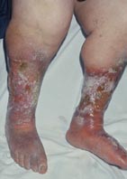 The
picture right shows ulceration with eczema and inverted champagne bottle
appearance.
The
picture right shows ulceration with eczema and inverted champagne bottle
appearance.
Abnormal appearance to the shape of the leg
(inverted champagne bottle)
An inverted champagne bottle aptly describes the appearances of some legs
with chronic venous insufficiency. The leg is very narrow at the ankle and
just above, but then becomes much fatter in the upper part of the calf below the
knee. This is commonly associated with pigmentation around the ankle and
sometimes with varicose veins.
Swelling
Swelling around the ankle, foot and lower leg especially of a mild degree can occur in many patients with venous
problems. If it becomes more severe and is only present in one leg, then it can be a sign that
investigation and treatment of the venous system is required.
Mention of an ulcer often concerns patients. An ulcer is simply an area that has lost the covering layer of skin so that the tissues beneath the skin are exposed. This is all that is meant by an ulcer. It does not say anything about the cause of the ulcer or how it will respond to treatment.
There is an important distinction between an ulcer and a graze on the leg. In the case of a graze only the superficial layers of the skin are lost even though this can be deep enough to cause bleeding. In an ulcer the whole thickness of the skin is lost and there are no skin cells in the defect. This difference has important implications for healing. In the case of a graze healing can take place over the whole graze, as there are still skin cells over the whole area. Healing is quick (5-10 days) because of these cells. In an ulcer the only way skin cells can bridge the ulcer and heal over is for the cells to grow in from the edges. This is a much slower process even in perfect conditions..
Leg ulcers are caused by two main problems in developed countries. The two commonest causes of ulceration are diseases of the veins and diseases of the arteries. As many as 75% of patients have a significant venous component to their leg ulcers. Arterial ulceration and mixed arterio-venous ulcers (ulcers due to a combination of venous and arterial disease) constitute the second major group of leg ulceration patients (14%). Diabetes mellitus can also cause ulceration, but predominantly in the foot. Venous and arterial problems can also occur in patients with diabetes.
| Causes of leg ulcers |
|---|
Post traumatic |
Vascular |
| Lymphatic |
| Vasculitic Rheumatoid arthritis Systemic lupus erythematosus Allergy Pyoderma gangrenosum |
| Metabolic Diabetes mellitus Necrobiosis lipoidica diabeticorum Gout |
| Haematological Polycythaemia rubra vera Leukaemia Sickle cell anaemia Thalassaemia Spherocytosis |
| Malignant Basal Cell Carcinoma Squamous cell carcinoma Melanoma Lymphoma |
| Infectious Bacterial Tubercular Fungal Syphilitic |
| Miscellaneous Drug induced eg hydroxyurea Self inflicted Post-irradiation Frostbite Insect bites |
| This is not an exhaustive list of causes of leg ulcers |
Sometimes ulcers can be due to skin cancers, although the majority of ulcers on the legs are not skin cancers. Rarely, a longstanding leg ulcer may develop into a skin cancer, usually a squamous cell carcinoma, commonly known as a Marjolin's ulcer. The table on the right gives a list of possible causes for leg ulcers. It is not an exhaustive list and many are very uncommon and not all universally recognised as true causes eg hypertensive ulceration. If you are aware of other causes of ulceration please e-mail me at [email protected] and I will be happy to add them to the list. If you images of rare types of ulcers I can also post them.
What sort of assessment do I need?
An accurate history and physical examination with special reference to venous and arterial disease, diabetes and rheumatoid arthritis is performed in all patients.
A formal assessment of the arterial circulation using the hand held Doppler and measurement of the ankle-brachial index is essential before instituting treatment.
Duplex scan of the venous system will clearly identify patterns of venous reflux which can be surgically corrected in appropriate patients.
History
Information about how the ulcer developed and progressed is important.
Many patients find their ulcer starts after a very minor injury which normally
would be expected to heal. Because of underlying disease in the arteries
or veins this doesn't happen and the ulcer deteriorates. Ulcers are less
likely to be successfully treated if they have been present for a long time or
if they are particularly large.
Varicose veins and previous deep venous thrombosis are possible contributing factors to the development of an ulcer. Previous surgery to the veins may be important in planning treatment. Your doctor will also ask about possible arterial problems in the legs. If you have suffered from intermittent claudication, previous ulcers or if you have had previous arterial bypass surgery to the legs, this will all be important. Your doctor will also ask about your general health and your mobility.
Examination
The examination will focus on the ulcer itself and on the arterial and
venous systems in the leg. The site and size of the ulcer, the edge of the
ulcer and the base of the ulcer are particularly important in deciding what sort
of ulcer is present. The appearance of the edge of the ulcer can raise suspicions of a possible skin cancer, but it also can indicate whether the ulcer is beginning to heal or if it is deteriorating. Very specific changes, such as a violaceous border, can indicate the presence of rare conditions such as pyoderma gangrenosum. The base of the ulcer can indicate if the ulcer will heal. Exposed bone or tendon, the presence of dead tissue or alternatively healthy granulation tissue are all important. Discharge, especially if smelly, can indicate the presence of infection. Discharge may be secondary to swelling in the leg. Constant discharge of fluid across the ulcer bed will impede healing.
Obvious varicose veins will be recorded. The pulses will be felt throughout the leg and the arterial circulation further assessed through colour and warmth of the limb.
Investigations
In all patients the blood pressure should be measured at the ankle using the
hand
held Doppler. This instrument is a very sensitive tool for assessing blood
flow in the leg, which is then compared against the blood flow in the arm. This
enables the ankle:brachial index (ABI) to be calculated. Usually the flow
of blood in the arms and legs is the same and the index is close to 1.0.
If there is impairment of the circulation in the lower limbs then the index will
be reduced to less than 1.0. The quality of the doppler signal is also important. A biphasic or triphasic signal will indicate good arterial flow even if the ABI is difficult to measure or pulses cannot be felt. It does need experience to be able to evaluate the doppler accurately.
Hand Held Doppler can also be used in the clinic to assess the veins, but the assessment is not as accurate as the that in the arteries. All patients require a more detailed assessment of the veins using colour flow ultrasound. This test supplies detailed information about the anatomy of the system of veins and the direction of blood flow in the veins.
If there is anything unusual about the appearance of the ulcer, your surgeon may suggest that a small piece of tissue is removed (biopsy). This will check whether there is any underlying skin cancer and may also help with other diagnoses in certain situations.
Yes, leg ulcers can be treated but the best form of treatment for your leg ulcer will depend on exactly why the ulcer has occurred.
Venous ulcers
Some ulcers are clearly caused by problems in the veins. This should
be confirmed by clinical assessment and on special investigations. If this
is the case then compression treatment should be commenced. It should only
be applied after the arteries have been assessed by measuring the ankle-brachial
index. This is because if compression is applied and the arteries are
badly diseased, this can damage the ulcer and the leg, and make matters worse.
It would also be very painful.
Before compression is applied, the leg and the ulcer should be thoroughly cleaned and a simple dry, non-adherent dressing applied to the ulcer itself. The ankle circumference is then measured and the compression system selected. The formal compression bandage is applied by a trained practitioner, usually a nurse skilled in bandaging techniques. The first layer consists of a soft wool bandage to protect bony points at the ankle and the shin bone. A crepe bandage is applied as the second layer. The third layer is an elasticated bandage that will apply compression. The final fourth layer applies further compression and keeps all of the bandages in place. Although this sounds quite complicated it is quite straightforward to apply in practice, by properly trained personnel. These bandages may be left in place for up to 7 days, but should be changed if fluid from the ulcer soaks through the bandages. If possible they are left, as it is thought that each dressing change damages some of the ulcer tissue that is trying to heal. This system is known as 4 layer compression (see opposite) and can be tailored to a certain extent to suit the particular shape and size of leg. In this link a nurse explains some of the features and treatment required for leg ulcers.
 Using these techniques it has been shown
that 50-70% of pure venous ulcers heal within 12 weeks. A recent randomised
trial has compared 4 layer bandaging with conventional ulcer treatments.
There was a significant improvement in healing in the 4 layer bandaging group
with 54% of ulcers healed at 3 months compared with only 34% in the control
group without compression (O'Brien JF et al, 2003). Others may heal
after this time depending on their size. If the ulcer fails to respond or the
patient is unable to initially tolerate compression then a period of bedrest and
elevation of the leg in
hospital can be helpful. This usually requires a stay of some weeks.
Bedrest helps by reducing swelling in the leg and therefore the amount of fluid
passing across the ulcer bed. Compression can usually then be applied to
maintain the effect when the patient is discharged.
Using these techniques it has been shown
that 50-70% of pure venous ulcers heal within 12 weeks. A recent randomised
trial has compared 4 layer bandaging with conventional ulcer treatments.
There was a significant improvement in healing in the 4 layer bandaging group
with 54% of ulcers healed at 3 months compared with only 34% in the control
group without compression (O'Brien JF et al, 2003). Others may heal
after this time depending on their size. If the ulcer fails to respond or the
patient is unable to initially tolerate compression then a period of bedrest and
elevation of the leg in
hospital can be helpful. This usually requires a stay of some weeks.
Bedrest helps by reducing swelling in the leg and therefore the amount of fluid
passing across the ulcer bed. Compression can usually then be applied to
maintain the effect when the patient is discharged.
Although the 4 layer system is one of the most common and effective in modern use, there are alternatives. A recent trial from St Thomas' Hospital in London compared 3 layer paste bandages with the conventional 4 layer system (Meyer FJ et al, 2003). In this study the 3 layer system was found to be more effective at healing than the 4 layer system. At the very least it is probably comparable and could certainly be used effectively in patients with intolerance to the standard 4 layer regime
Despite intensive treatment there will still be some patients who are left with intractable ulcers that fail to heal. In these circumstances the aim of treatment is to keep the ulcer under control and reduce its' effect on the day to day life of the patient as much as possible.
Arterial ulcers
Some ulcers are mainly arterial even if they have a venous component.
This is always the case when the ABI is less than 0.5, indicating a severe
degree of arterial impairment. In these circumstances compression should
never be applied. These ulcers should be managed by examining the arteries
in more detail by ultrasound and angiography.
Usually patients with these ulcers will require some treatment to improve the
blood supply if their ulcer is going to heal.
Mixed ulcers
In some patients the ulcers are caused by a combination of problems in the
arteries and the veins. It can sometimes be difficult in these patients to
decide the most effective way of managing their ulcer. In general if the
Ankle-Brachial Index is greater than 0.5, but less than 0.8, it is often sensible to try modified
(lighter) compression as a first option. If this is tolerated and appears
to be helping the ulcer to heal, then it should be continued. If it is not
tolerated or appears to be unhelpful then it will be important to investigate
the arteries in the same way as for arterial ulcers (Humphreys
ML, 2007).
General
Although ulcers may have predominantly venous or arterial components, there are usually many factors that contribute to the development of an ulcer. The common factors are obesity, immobility, ankle and leg swelling from other causes and poor ankle movements.
Ulcer dressings and antibiotics
In general provided the blood supply to the
tissues is good and compression is applied, if appropriate, it hardly matters
which dressing is placed onto the ulcer bed. As long as it is clean, dry
and non-adherent the ulcer should respond. Many claims are made for
different types of dressings, most of which are hard to substantiate.
Regular changes of dressing type usually have little value and may
actually do harm as patients often develop allergic reactions (dermatitis).  A recent review and meta-analysis of dressings for venous ulcers showed that the
type of dressing applied beneath compression was not shown to influence healing
(Palfreyman S, Nelson EA,
Michaels JA, 2007).
A recent review and meta-analysis of dressings for venous ulcers showed that the
type of dressing applied beneath compression was not shown to influence healing
(Palfreyman S, Nelson EA,
Michaels JA, 2007).
Moist wound healing has become the mantra amongst wound "specialists". There is probably nothing fundamentally wrong with moist wound healing but the evidence supporting this approach is not good. For more information click here.
Antibiotics are also frequently prescribed for ulcers on the basis of a swab result that has grown bacteria. Antibiotics should only be used when there is frank infection. This usually means a hot, red, tender leg. Because we are all covered with bacteria a swab taken from anywhere on the body whether ulcerated or not will grow bacteria, but they do not require treatment. They are a normal part of the body flora and the presence of these normal bacteria is known as colonisation. Overtreatment with antibiotics is likely to lead to problems with antibiotic resistance developing in bacteria that are present, making the future treatment of infection even more difficult.
There are many dressings popularly thought to aid ulcer healing. Manuka honey has recently bee subject to a randomised trial in which 368 patients with leg ulcers were randomised to either conventional care or dressing impregnated with manuka honey. Honey dressings did not improve venous ulcer healing at 12 weeks (Jull et al 2008). In New Zealand Kawa-kawa leaves are also thought to be of benefit but there is no scientific evidence to support their use.
Cleaning your leg
It is perfectly acceptable to clean your leg and ulcer with ordinary tapwater. There is no benefit in using sterile water or saline. If you have a planned visit from the district nurse then, by arrangement, it can be useful to shower and clean the leg, if your are able, prior to the planned visit. Only do this after discussion.
What is the place of venous surgery?
In patients with venous ulcers (ulcers caused by venous disease only) there is abnormal reverse flow (reflux) in the veins. Colour flow Duplex ultrasound scanning can accurately identify the sites of reflux in the majority of patients. The scan is a painless procedure which takes about 30 minutes for one leg. Following a scan there are 4 main categories of results:
1. Superficial venous reflux only - this situation is correctable by surgery. There is good evidence that performing surgery to correct the reflux will reduce the risk of recurrence (see ESCHAR study below).
2. Deep venous reflux only - this situation is not correctable by surgery and the mainstay of treatment is continued compression. Operations to repair the valves in the veins have been devised, but are only performed by a few surgeons worldwide. Although early results often appear good in the hands of individuals, they are often difficult to reproduce. Newer techniques to implant valves using endovascular techniques will need thorough investigation before they can be recommended.
3. Mixed superficial and deep venous reflux - the ESCHAR study has now confirmed that with longer follow up surgery to correct the reflux in the superficial veins will benefit these patients. The surgery can easily be performed, but even if successful in eliminating superficial reflux will still leave the patient with uncorrectable deep venous reflux.
4. Occluded deep venous system - sometimes after a deep venous thrombosis the vein will not be re-opened by the repair mechanisms of the body and will remain blocked. When this happens the superficial veins can become enlarged to compensate and carry blood back to the heart. If this is the case the superficial varicose veins should not be removed except after detailed assessment under a specialised vascular surgeon.
These are the main categories, although there are many different possible combinations and detailed discussion of the surgical options should take place with your specialist surgeon. It is important to remember that in patients with ulcers the indication for surgery is to reduce the risk of further ulceration and to facilitate healing of pre-existing ulcers by eliminating reflux in the veins. Cosmetic benefits may also be apparent, but this is not the primary aim of surgery.
The ESCHAR study (Gohel MS, Barwell JR, Taylor M et al.), has reported final results on healing and recurrence rates after treatment with compression with or without the addition of surgery to the veins in people with venous leg ulcers. Leg ulcer healing rates at 3 years were 89% for the compression group and 93% for the compression plus surgery group. Rates of ulcer recurrence at 4 years were 56% for the compression group and 31% for the compression plus surgery group. For patients with isolated superficial venous reflux, recurrence rates at 4 years were 51% for compression and only 27% for compression plus surgery. Results were similar in patients with superficial and segmental deep venous reflux (52% versus 24%) at three years . In patients with superficial and total deep venous reflux, there was still benefit from superficial venous surgery with leg ulcer recurrence rates improved from 46% with compression alone, to 32% with the addition of surgery. The authors concluded that most patients with chronic venous ulceration will benefit from the addition of simple venous surgery to reduce ulcer recurrence rates.
In the study overall, 40% of patients refused to undergo venous surgery and this is a reflection of the elderly and infirm population that often presents with leg ulceration. Less invasive treatments now available may offer benefits to this group of patients.
It is possible to develop further leg ulcers and the risk varies from study to study. To reduce your risk of further ulcers it is likely to be necessary to wear graduated compression stockings. These stockings provide maximum compression at the ankle and this gradually reduces up the leg. Precise measurements of the leg are required to fit the stockings correctly and 2 pairs should be provided so that one can be washed at any one time. The stockings should be replaced approximately every 6 months. Probably between 5-30% of patients will develop further leg ulceration in the first 2 years after ulcer healing.
Once ulcers have healed Class 2 below knee compression stockings should be worn. Compliance with wearing stockings is notoriously poor except where dedicated nurses review patients regularly. In patients who wear their stockings regularly, recurrent ulceration can be reduced to 32% at 5 years from 69% in patients who do not wear their stocking.
The majority of patients with leg ulcers can be managed in the community by competently trained nurses working to set protocols. The most appropriate model of care for these patients appears to be one consisting of a series of strategically placed community leg ulcer clinics staffed by suitably trained nurses and supported by vascular surgery. Chronic leg ulcer is perfectly suited to shared care with a blend of hospital based specialist intervention and community based care.
Leg ulcer clinics based in the community have been shown in a UK study to provide more clinically effective and cost effective care when compared with traditional home based care delivered by district nurses. New Zealand, however, is not the UK and has a significant rural population often long distances from the nearest town. Despite this there is no doubt that patients report better dressings and improved outcomes from a group of nurses who perform compression bandaging in specialist clinics. This is because they see the same patients frequently and are able to learn to optimise their bandaging technique to provide the best outcome. Dedicated follow up by the same nurses achieves the best results. Too often patients report that they never have the same nurse twice to do the dressing and consequently it is difficult for those nurses to develop their technique in isolation with no feedback.
![]() Bookmark this on Delicious
Bookmark this on Delicious  reddit
![]() facebook
facebook
Bergan JJ et al.
Chronic venous disease N Engl J Med 2006; 355: 488-98.
Criqui MH et al. Risk factors for chronic venous disease: The San Diego population study. J Vasc Surg 2007; 46: 331-337.
Douglas WS, Simpson NB. Guidelines for the
management of chronic venous leg ulceration. Report of a multidisciplinary
workshop. Brit J Dermatol 1995; 132: 446-452.
Grabs AJ, Wakely MC, Nyameke I,
Ghauri ASK, Poskitt KR. Colour duplex ultrasonography in the rational management
of chronic venous leg ulcers. Brit J Surg 1996; 83: 1380-1382.
Scriven JM, London NJM. Single-visit
venous ulcer assessment clinic: the first year. Brit J Surg 1997; 84: 334-336.
Ghauri ASK, Poskitt KR. Single-visit
venous ulcer assessment clinic: the first year. Brit J Surg 1997; 84: 1323.
Fletcher A, Cullum N, Sheldon TA. A
systematic review of compression treatment for venous leg ulcers. Brit Med J
1997; 315: 576-580.
Blair SD, Wright DDI, Backhouse CM,
Riddle E, McCollum CN. Sustained compression and healing of chronic venous
ulcers. Brit Med J 1988; 297: 1159-1161.
Anonymous. Compression therapy for
venous leg ulcers. Drug and Therapeutics Bulletin 2000; 38(4): 28-31.
NHS Centre for Reviews and
Dissemination. Compression therapy for venous leg ulcers. Effective healthcare
1997; 3(4): 2-12.
Bello M, Scriven M, Hartshorne T,
Bell PRF, Naylor AR, London NJM. Role of superficial venous surgery in the
treatment of venous ulceration. Brit J Surg 1999; 86: 755-759.
Harper DR, Nelson EA, Gibson B,
Prescott RJ, Ruckley CV. Prevention of recurrence of venous ulceration:
prospective randomised controlled trial over 5 years of class 2 and class 3
elastic compression. Phlebology 1995; 2(suppl 1): 872-873.
Moffatt CJ, Dorman MC. Recurrence of
leg ulcers within a community ulcer service. J Wound Care 1995; 4: 57-61.
Ruckley CV. Caring for patients with
chronic leg ulcer. Brit Med J 1998; 316: 407-408.
Morrell CJ, Walters SJ, Dixon S et
al. Cost effectiveness of community leg ulcer clinics: randomised controlled
trial. Brit Med J 1998; 316: 1487-1491.
Moffat CJ, Franks PJ, Oldroyd M et
al. Community clinics for leg ulcers and impact on healing. Mrit Med J 1992;
305: 13891392.
Bosanquet N, Franks P, Moffat C et
al. Community leg ulcer clinics: cost-effectiveness. Health Trends 1993; 25(4):
146-148.
Simon DA, Freak L, Kinsella A et al.
Community leg ulcer clinics: a comparative study in two health authorities. Brit
Med J 1996; 312: 1648-1651.
Anonymous. Dressings for leg ulcers.
Drugs and Therapeutics Bulletin. 1986; 24(3): 9-12.
Scottish Intercollegiate Guidelines
Network. The care of patients with chronic leg ulcer. A national clinical
guideline. SIGN publication No26; 1998.
O'Brien JF, Grace PA, Perry IJ et al. Randomised clinical trial and economic
analysis of four layer compression bandaging for venous ulcers. Brit J Surg
2003; 90: 794-798.
Meyer
FJ, McGuiness CL, Lagattolla NRF, Eastham D, Burnand KG. Randomised clinical
trial of three-layer paste and four-layer bandages for venous leg ulcers. Brit J
Surg 2003; 90: 934-940.
Humphreys ML, Stewart AHR,
Gohel MS et al. Management of mixed arterial and venous ulcers. Brit J
Surg 2007; 94: 1104-07.
Palfreyman S, Nelson EA,
Michaels JA. Dressings for venous leg ulcers: systematic review and
meta-analysis. Brit Med J 2007; 335: 244.
Jull A, Walker N, Parag V et al. Randomised clinical trial of honey-impregnated dressings for venous leg ulcers. Brit J Surg 2008; 95: 175-82.
Gohel MS, Barwell JR, Taylor M
et al. Long term results of compression therapy alone versus
compression plus surgery in
chronic venous ulceration (ESCHAR study): randomised controlled trial. Brit Med
J 2007; 335: 83-89.
Last updated Saturday, 6 November, 2010 8:48 PM